|
Email / Resume / Github / Google Scholar I am a graduate student in Electrical
Engineering and Computer Science at the Massachusetts
Institute of Technology. I received my undergraduate degree at the
University of California, Berkeley. I am interested in optimization theory, functional analysis, computational imaging, image processing, computer vision and machine learning. My research thus far has been primarily focused on the design and consequent solving of linear inverse compressed sensing problems in Magnetic Resonance Imaging. I am currently advised by Kawin Setsompop. At UC Berkeley, I worked under the guidance of Frank Ong, Jon Tamir and Michael Lustig. |
|
|
|
|
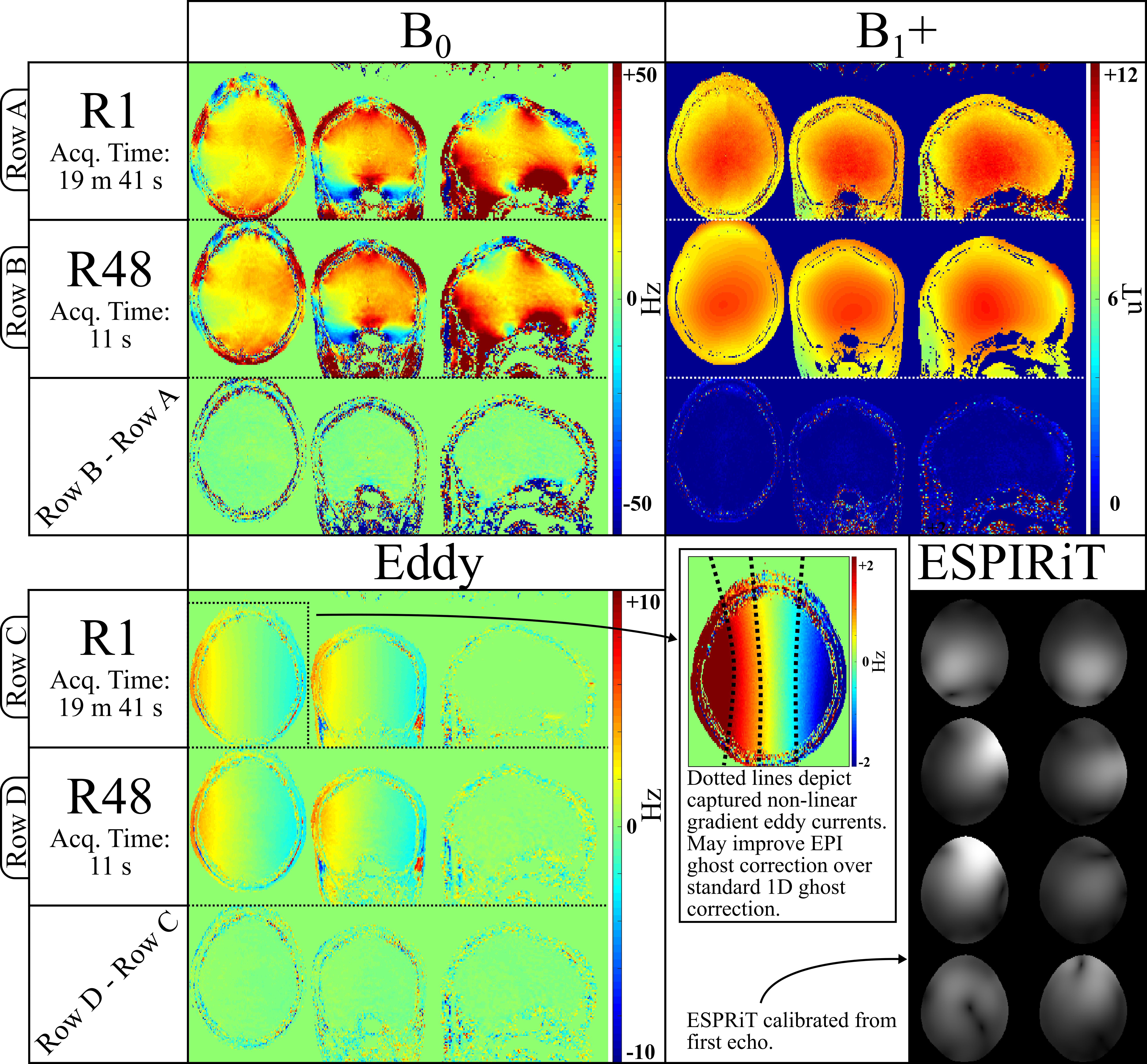
Abstract. Calibration scans that acquire coil sensitivity, B0, and B1+ inhomogeneities information play an important role in enabling modern acquisition and reconstruction techniques. This work proposes a unified, rapid calibration sequence termed Physics Calibration (PhysiCal) to obtain accurate B0, Eddy, B1+ and coil sensitivity maps. PhysiCal utilizes a carefully designed mix of full and variable density sampling acquisitions across echoes with synergistic constrained and eigenvalue reconstruction for robust and accurate recovery of whole-brain B0, B1+, Eddy and 32-channel coil sensitivity maps in just 11 seconds at 1 mm x 2 mm x 2mm resolution at 3T. Summa Cum Laude awarded. Links: Abstract / Talk coming soon. |
|
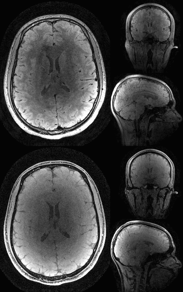
Abstract. Previously, we presented Wave-Shuffling: a technique that combines random-sampling and the temporal low-rank priors of Shuffling with the sinusoidal gradients of Wave-CAIPI to achieve highly-accelerated, time-resolved acquisitions. In this work, we optimize and apply Wave-Shuffling to T2-SPACE and MPRAGE to achieve rapid, 1 mm-isotropic resolution, time-resolved imaging of the brain where we recover a time-series of clinical contrast. We also present preliminary explorations into Joint-Contrast Wave-Shuffling to boost signal-to-noise ratio and reconstruction-conditioning at high accelerations. Magma Cum Laude awarded. |
|
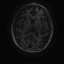
Abstract. T2-shuffling is a recently proposed approach that can reconstruct multiple, sharp T2-weighted images from a single fast spin-echo (FSE) scan. Wave-CAIPI is a parallel imaging technique that uses additional sinusoidal gradients to spread aliasing in the readout direction, to take full advantage of coil-sensitivity information in highly accelerated rectilinear acquisitions. In this work, we augment T2-shuffling with wave-encoding and examine the ability of this combined approach in accelerating FSE acquisition that can achieve multiple T2-weighted images reconstruction. We demonstrate the efficacy of our technique through a 2D simulation, where wave-encoding was shown to provide good reconstruction at 2-3x higher accelerations. |
|
Abstract. ESPIRiT is a robust, auto-calibrating approach to parallel MR image reconstruction that estimates the subspace of sensitivity maps using an eigenvalue-based method. While it is robust to a range of parameter choices, having parameters that result in a tight subspace yields the best performance. We propose an automatic, parameter free method that appropriately weights the subspace using a shrinkage operator derived from Stein's Unbiased Risk Estimate. We demonstrate the efficacy of our technique by showing superior map estimation without user intervention in simulation and in-vivo data compared to the current default method of subspace estimation. Summa Cum Laude awarded. Links: Abstract / Talk / Preprint / Publication |
|
Abstract. MRI is a safe and powerful tool that can be used to image both anatomy and function. Acquiring 3D MRI over time has several clinical applications:
Link: Poster |
|
|
|
University of California, Berkeley
University of California, Berkeley
|
|
Website template modified from Jon Barron. |