High-Content Digital Pathology

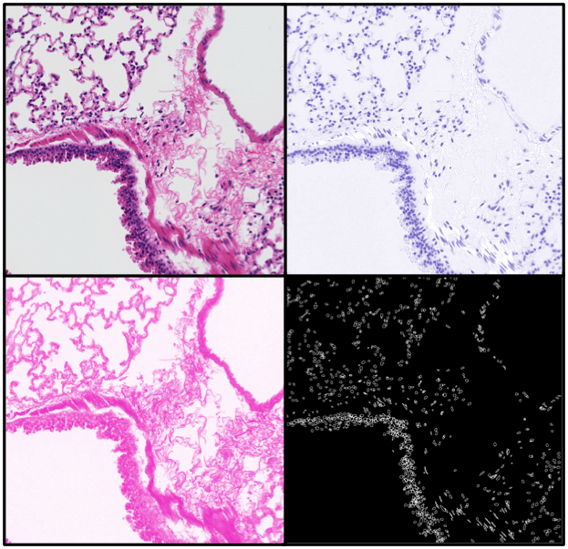
We analyze large amounts of histological data digitally for thorough investigation of tissues and statistical insight. Algorithms have been designed to characterize cellular status and functionality. Spectral separation methods provide more contrast for identifying key features such as nuclei, cell boundaries, shape, contours and inclusions. Additional image processing and Computer Vision techniques are used for feature detection and classification which aid in the pathological categorization for histology of the heart, lung, liver, etc.
Similar techniques may be applied in the near infrared, where hardware provides extended spectral resolution.