Commercial Medical
Microscopy Development
Commercial Medical
Microscopy Development

The Cytyc Corporation ThinPrep® Imaging System (Imager) is a device that uses computer imaging technology to assist in primary cervical cancer screening of ThinPrep Pap Test slides for the presence of atypical cells, cervical neoplasia, including its precursor lesions (Low Grade Squamous Intraepithelial Lesions, High Grade Squamous Intraepithelial Lesions), and carcinoma as well as all other cytologic criteria as defined by 2001 Bethesda System: Terminology for Reporting Results of Cervical Cytology.
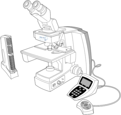
The ThinPrep Imaging System is an automated imaging and review system for use with ThinPrep Pap Test slides. It combines imaging technology to identify microscopic fields of diagnostic interest with automated stage movement of a microscope in order to locate these fields. In routine use, the ThinPrep Imaging System selects 22 fields of view for a Cytotechnologist to review. Following review of these fields, the Cytotechnologist will either complete the diagnosis if no abnormalities are identified or review the entire slide if any abnormalities are identified. The ThinPrep Imaging System also allows the physical marking of locations of interest for the Cytopathologist.
The ThinPrep Imaging System consists of an Image Processor and one, or more, Review Scopes. The system makes use of computer imaging to select fields of view for presentation to a Cytotechnologist on a Review Scope. Slides used with this system must first be prepared on a ThinPrep 2000 or 3000 Processor, and stained with ThinPrep Stain.
The Imaging Processor acquires and processes image data from the slides to identify diagnostically relevant cells or cell groups based on an imaging algorithm that considers cellular features and nuclear darkness. During slide imaging, the alphanumeric slide accession identifier is recorded and the x and y coordinates of 22 fields of interest are stored in the computer database. This computer also coordinates the communication of information between the Image Processor and the Review Scopes.
ThinPrep® Imaging System
Automated White Blood Cell Differential
The white blood cell differential assesses the ability of the body to respond to infection and eliminate it. The results indicate the severity of allergic and drug reactions plus the response to parasitic and other types of infection. The process of cell differentiation is essential in evaluating the reaction to viral infections and response to chemotherapy. Furthermore, it indicates the various stages of leukemia.
A precise multi-level platform separates white blood cells from red blood cells. Computer vision techniques, such as ellipse fitting and feature detection, are used to identify and classify the white blood cells as Neutrophils, Eosinophils, Basophils, Lymphocytes, or Monocytes.
2000-2001, Battelle Memorial Institute, Product Development Group