Functional Imaging of Neurons

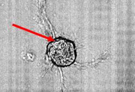
We demonstrate how optical coherence imaging techniques can detect intrinsic scattering changes that occur during action potentials in single neurons. Using optical coherence tomography (OCT), an increase in scattering intensity from neurons in the abdominal ganglion of Aplysia californica is observed following electrical stimulation of the connective nerve. In addition, optical coherence microscopy (OCM), with its superior transverse spatial resolution, is used to demonstrate a direct correlation between scattering intensity changes and membrane voltage in single cultured Aplysia bag cell neurons during evoked action potentials. While intrinsic scattering changes are small, OCT and OCM have potential use as tools in neuroscience research for non-invasive and non-contact measurement of neural activity without electrodes or fluorescent dyes. These techniques have many attractive features such as high sensitivity and deep imaging penetration depth, as well as high temporal and spatial resolution. This study demonstrates the first use of OCT and OCM to detect functionally-correlated optical scattering changes in single neurons.
Neural Imaging