Developmental Biology

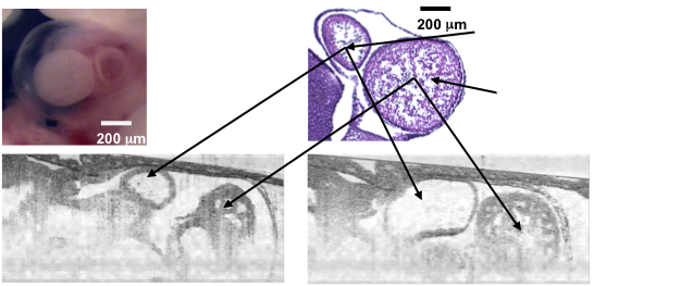
Optical coherence tomography (OCT) is an emerging high-resolution real-time biomedical imaging technology that has potential as a novel investigational tool in developmental biology and functional genomics. In this study, murine embryos and embryonic hearts are visualized with an OCT system capable of 2-micron axial and 15-micron lateral resolution and with real-time acquisition rates. We present, to our knowledge, the first sets of high-resolution 2- and 3-D OCT images that reveal the internal structures of the mammalian (murine) embryo and embryonic cardiovascular system. Strong correlations are observed between OCT images and corresponding hematoxylin- and eosin-stained histological sections. Real-time in vivo embryonic heart activity is captured by spectral-domain optical coherence tomography, processed, and displayed at a continuous rate of five frames per second. With the ability to obtain not only high-resolution anatomical data but also functional information during cardiovascular development, the OCT technology has the potential to visualize and quantify changes in murine development and in congenital and induced heart disease, as well as enable a wide range of basic in vitro and in vivo research studies in functional genomics.
Murine Cardiac Development
