 The realization that the structure of a protein determines its function was a profound revelation to me
as an undergraduate Biochemistry major at Lebanon Valley College.
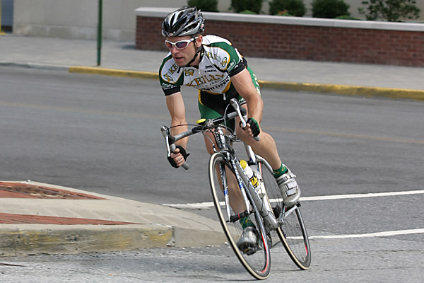
This idea is so fundamental that it's akin to stating, "the structure of a wheel allows it to roll."
Knowing the structure of our bicycle wheel allows us to understand why a broken spoke might make the wheel lopsided
and explain why we crashed our bike. Similarly, understanding protein structure is essential, not just because it's the basis
for life, but because knowledge of structure allows us to understand how
defects cause disease and to develop treatments.
The realization that the structure of a protein determines its function was a profound revelation to me
as an undergraduate Biochemistry major at Lebanon Valley College.
This idea is so fundamental that it's akin to stating, "the structure of a wheel allows it to roll."
Knowing the structure of our bicycle wheel allows us to understand why a broken spoke might make the wheel lopsided
and explain why we crashed our bike. Similarly, understanding protein structure is essential, not just because it's the basis
for life, but because knowledge of structure allows us to understand how
defects cause disease and to develop treatments.
Proteins are not easily seen or understood. To learn some of the skills needed to study protein structure and function, I entered the Biochemistry, Cellular and Molecular Biology program at Johns Hopkins University, School of Medicine where I worked in the lab of Wade Gibson. In the Gibson lab I studied an extremely challenging protein: a protease (protein that eats other proteins) whose substrate (favorite snack) was itself. You can only imagine what it’s like trying to work with something that eats itself! This particular protease is produced by cytomegalovirus (CMV) and is essential for assembly of an infectious virus particle. It remains an untapped target for treatment of CMV infection.
 After taming this wild protease to elucidate some aspects of its catalytic activity,
ability to self-interact, and function in mutant viruses, I wanted to learn how to
"see" proteins. Some proteins are well behaved, and like our bicycle wheel
can be readily defined structurally. Other proteins are assembled from many
moving parts, like a cyclist on a bicycle. Large, dynamic macromolecules can be directly
visualized using electron microscopy. I was fortunate to study the art of
single-particle electron microscopy at
The Scripps Research Institute with
Francisco Asturias,
who has applied the method to some very complicated, structurally heterogeneous proteins.
My studies on mammalian fatty acid synthase revealed flexibility in the structure
that explained some controversial functional observations made by our collaborators in
Stuart
Smith’s lab. I also collaborated with
Roger Kornberg’s lab at Stanford to visualize transcription factor II H,
a 10-component protein responsible for unwinding DNA at the start of a gene,
allowing the gene to be transcribed or repaired when damaged.
After taming this wild protease to elucidate some aspects of its catalytic activity,
ability to self-interact, and function in mutant viruses, I wanted to learn how to
"see" proteins. Some proteins are well behaved, and like our bicycle wheel
can be readily defined structurally. Other proteins are assembled from many
moving parts, like a cyclist on a bicycle. Large, dynamic macromolecules can be directly
visualized using electron microscopy. I was fortunate to study the art of
single-particle electron microscopy at
The Scripps Research Institute with
Francisco Asturias,
who has applied the method to some very complicated, structurally heterogeneous proteins.
My studies on mammalian fatty acid synthase revealed flexibility in the structure
that explained some controversial functional observations made by our collaborators in
Stuart
Smith’s lab. I also collaborated with
Roger Kornberg’s lab at Stanford to visualize transcription factor II H,
a 10-component protein responsible for unwinding DNA at the start of a gene,
allowing the gene to be transcribed or repaired when damaged.
But electron microscopy is only one way to visualize protein structure. Proteins that are better defined structurally can sometimes be crystallized and visualized at higher resolution using X-rays. Cathy Drennan’s lab uses this method to understand how proteins catalyze difficult reactions. I recently joined her group to learn X-ray crystallography and to work closely with a multidisciplinary team that is throwing every imaginable method at ribonucleotide reductase in collaboration with the Asturias lab and JoAnne Stubbe’s lab. At every turn this amazing enzyme presents us with a new,unexpected surprise. Stay tuned …